

Importance of Mucus
Integrating mucus into in vitro models is essential to modelling a physiologically relevant system including digestive fluids, mucus and absorptive cells
What is mucus?
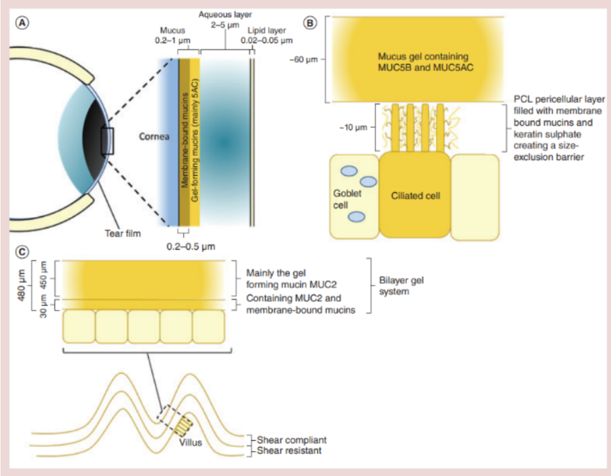
Mucus is a gel secreted by cells that coats various epithelial surfaces throughout the body, including the eye, airways, stomach, small intestine, colon and cervicovaginal tract. It's important in absorption and protection in these external surfaces of the body.
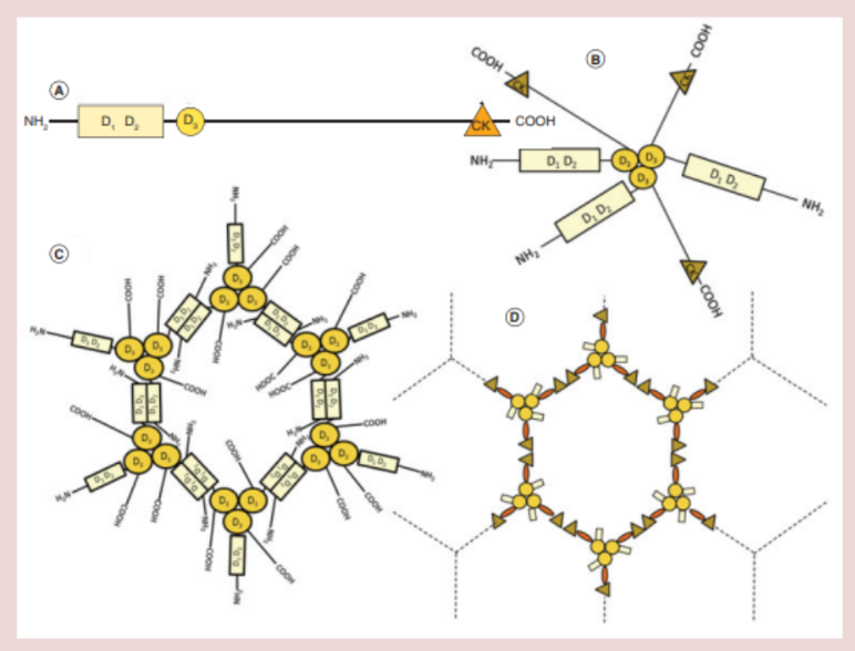
Mucus is a hydrogel and typically consists of around 95% water, but the major gel forming component is a glycoprotein molecule called mucin. Mucin molecules bond and interact with each other to form net-like gels with various functions depending on where they are secreted. As well as mucin, mucus contains proteins, lipids, DNA, and other shed cell debris.
The function of mucus in the digestive tract
The primary function of a surface mucus layer in the digestive tract is to protect the underlying epithelial tissues.
The digestive tract is lined with specialised but delicate epithelial layers, which make up a total surface area of around 32 m². These epithelia are under constant physical, chemical and biological stress, all of which are normal and crucial aspects of digestive tract function. Chewing, stomach churning and peristalsis, when combined with abrasive ingested substances, subject the delicate epithelia to high shear forces. Endogenous digestive secretions from the stomach and pancreas, including hydrochloric acid, bicarbonate, bile and a cocktail of digestive enzymes, have the potential to cause catastrophic damage to the epithelia. Ingested microorganisms, like H. pylori, also pose a significant threat through the secretion of dangerous toxins or direct tissue infection.
These stresses make the digestive tract an inhospitable environment for delicate tissues, but epithelial cells need to be in close contact with these digestive fluids in order to absorb nutrients from digestion. Evolution's solution to this problem is the surface mucus layer, which lines these epithelial surfaces and protects them from damage, while allowing the permeation of key nutrients.
Mucus in the small intestine
The small intestine is a digestive and absorptive and is the primary site of nutrient and drug absorption – therefore is of key importance for in vitro modelling.
The small intestinal mucus layer must be able to protect the underlying epithelium from digestive enzymes, bile and microorganisms, but also must be permeable enough to allow efficient nutrient absorption. The mucus layer achieves this by being selectively permeable. It has been shown that SI mucus has a pore size of 200-500 nm in diameter. This allows the mucus gel to act as a size exclusion barrier. Any molecules larger than this will not access the epithelium, while smaller molecules will access the epithelium and be available for brush border digestion & absorption.
The rapid turnover of mucus at the epithelial surface ensures that the mucus layer is not compromised by digestion by host enzymes or microbes.
The physiology of small intestinal mucus is incredibly important when considering the delivery of orally delivered products including nutraceuticals, and drugs. The pore size of small intestinal mucus may be the limiting factor to absorption if the active ingredient is larger than 200-500 nm in diameter, and some molecules with complex structures (e.g. long branching molecules) may get trapped between pores despite being small enough to fit through. Another barrier to absorption may by the charge of the mucins. The glycan chains of small intestinal mucus harbour terminal ester sulphates and sialic acid residues – these residues give the mucin polymers a negative charge. Target molecules with positive charges may interact with the negatively charged residues on mucins, becoming trapped. Negatively charged molecules may also be impacted, as they could be repelled from the mucins layer preventing permeation, or driven through the mucins layer.
In vitro modelling of intestinal absorption
There are many different techniques used to model small intestinal absorption in vitro. The industry standard is the Caco-2 cell permeability assay. Caco-2 cells are a cancer cell line that mimic the SI enterocyte phenotype when differentiated. The assay utilises a Caco-2 cell epithelial layer grown on a membrane which separates an upper and lower chamber. An active ingredient can be dosed to either side to estimate intestinal absorption or efflux. Similar techniques can be employed with more complex epithelial layers, such as primary intestinal epithelial cells, or multicellular epithelial layers derived from organoids.
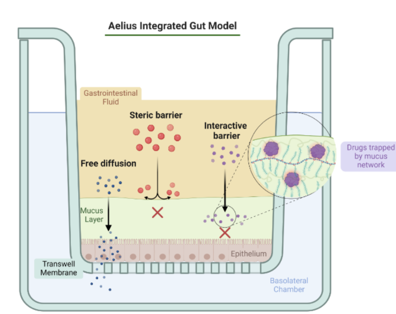
One thing that these models share is the lack of a protective surface mucus layer. Therefore, the luminal environment of the small intestine cannot be simulated as components like digestive enzymes and bile will cause significant damage to the epithelium. Any important interactions of the active ingredient and luminal contents will be excluded. The absence of a mucus permeation phase further limits the biological relevance of these systems, and without the protection of a mucus layer, testing formulations involving cosolvents and emulsifiers is not feasible.
Our Integrated Solution
At Aelius Biotech, our biocompatible and protective mucus gel is a solution to these problems. By using this gel in our Caco-2 assay, we can incorporate the digestive, mucus permeation and epithelial absorption phases, all in a single integrated system.
Bibliography
Experience the Difference
Discover how our unique mucus modelling technology can enhance your research and development outcomes
Speak to an Expert